Advanced Quantitative Fluorescence Microscopy to Probe the Molecular Dynamics of Viral Entry, Science Lab
Por um escritor misterioso
Descrição
Viral entry into the host cell requires the coordination of many cellular and viral proteins in a precise order. Modern microscopy techniques are now allowing researchers to investigate these interactions with higher spatiotemporal resolution than ever before. Here we present two examples from the field of HIV research that make use of an innovative quantitative imaging approach as well as cutting edge fluorescence lifetime-based confocal microscopy methods to gain novel insights into how HIV fuses to cell membranes and enters the cell.

Probing the Allosteric Inhibition Mechanism of a Spike Protein Using Molecular Dynamics Simulations and Active Compound Identifications

Phasor S-FLIM workflow and robustness to noise of the phasor

Influenza A virus exploits transferrin receptor recycling to enter host cells
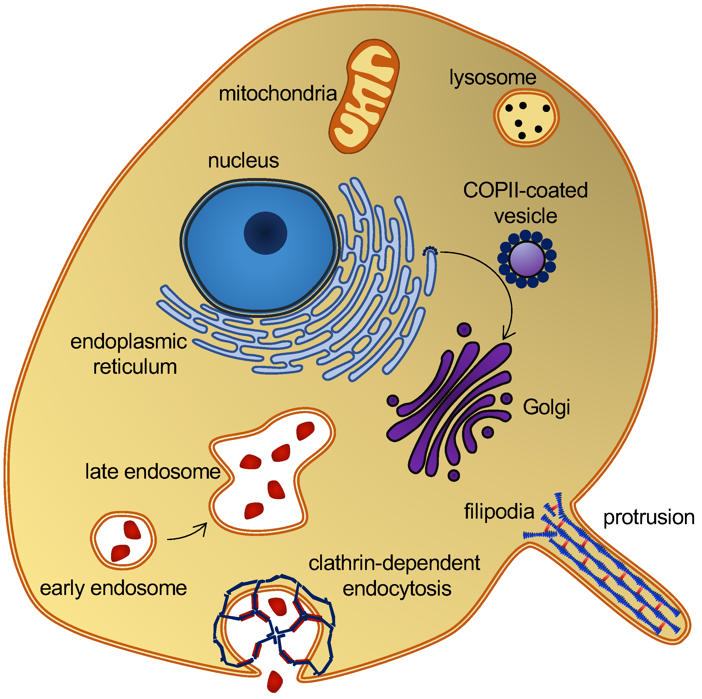
IJMS, Free Full-Text

Aptamers targeting SARS-COV-2: a promising tool to fight against COVID-19: Trends in Biotechnology

A spatial multi-scale fluorescence microscopy toolbox discloses entry checkpoints of SARS-CoV-2 variants in Vero E6 cells - Computational and Structural Biotechnology Journal

HIV-1 Vpr induces ciTRAN to prevent transcriptional repression of the provirus

Processes, Free Full-Text

NgR1 binding to reovirus reveals an unusual bivalent interaction and a new viral attachment protein

Primate-conserved carbonic anhydrase IV and murine-restricted LY6C1 enable blood-brain barrier crossing by engineered viral vectors

High-Speed AFM Reveals Molecular Dynamics of Human Influenza A Hemagglutinin and Its Interaction with Exosomes
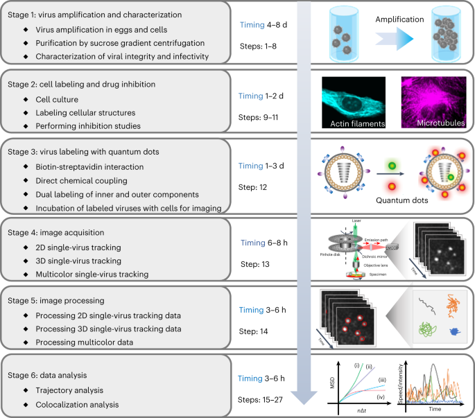
Single-virus tracking with quantum dots in live cells
Detection of fluorescently-labelled influenza virus using
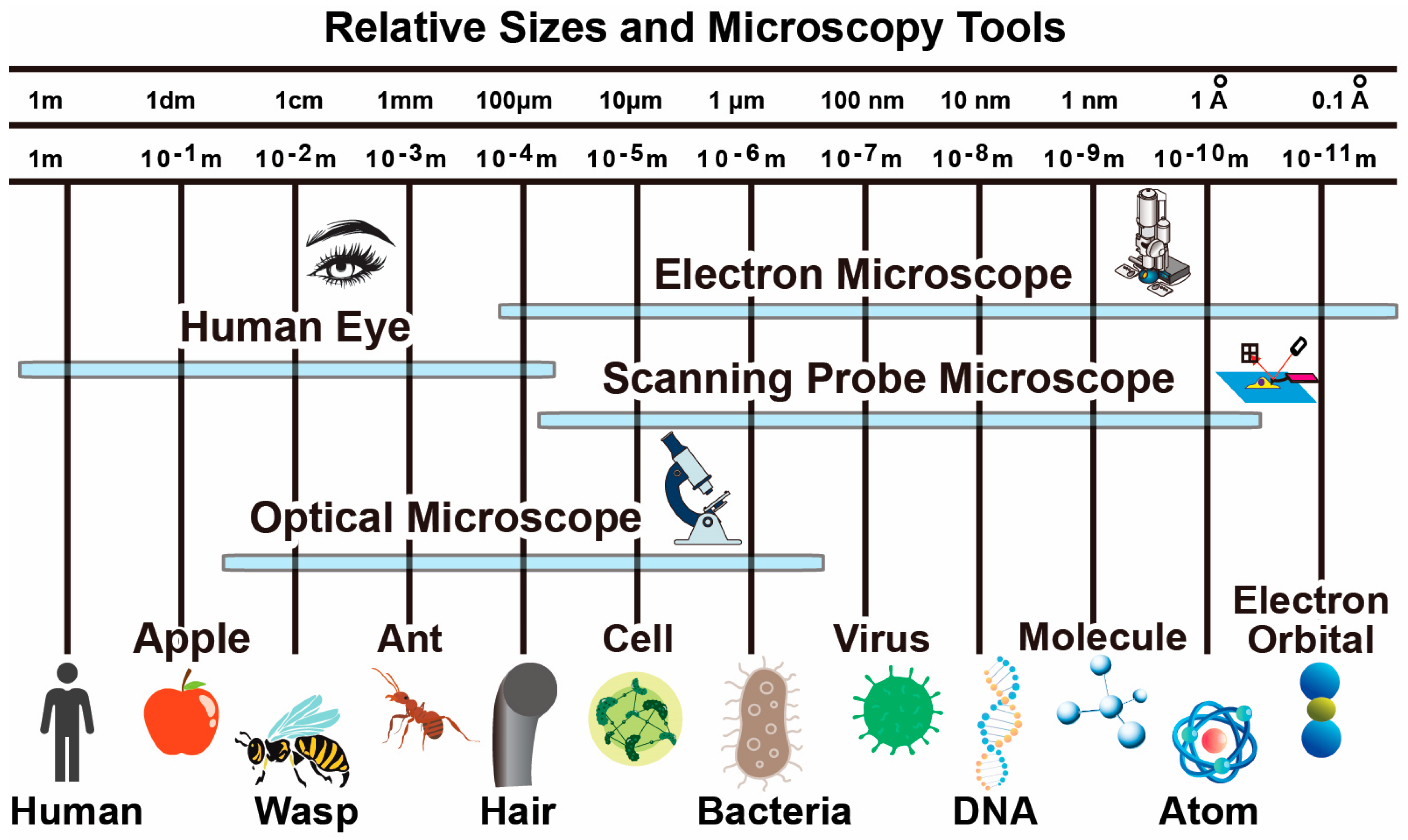
Biosensors, Free Full-Text
de
por adulto (o preço varia de acordo com o tamanho do grupo)


